Photosynthetic Sea Slug Pirates

We all know that plants ‘eat’ through the process called photosynthesis, a chemical process where light energy and carbon dioxide are turned into carbohydrates, oxygen and water.  In plants and algae, the process of photosynthesis occurs in the chloroplast, an organelle found within the individual cells.  It has become widely accepted that the plant and algal chloroplast originated as a cyanobacteria that was engulfed by another single celled organism, resulting in a symbiotic relationship. After this primary ‘endosymbiotic event’ the original cyanobacteria (now chloroplast) transferred genes that are needed for its survival to the nucleus of the symbiont.  The symbiont makes proteins needed to maintain the chloroplast, and in turns receives carbohydrates produced in the chloroplast for its own survival.

Illustration of the origin of the plant and algal chloroplast. Image from: http://biol10005.tumblr.com/post/32506576425/evolution-of-the-eukaryotic-cell

Plants and algae are not the only organisms that have found a way to photosynthesize.  A few species of sacoglossan sea slugs manage to steal the chloroplasts from some of the species of algae that they eat and are able to maintain these ‘kleptoplasts’ for their own benefit. It has been known that these sea slugs are able to maintain the kleptoplasts for extended periods of time, enabling the slugs to survive during extended periods of starvation.  However the mechanisms behind the longevity of the kleptoplasts remains elusive.  Normal photosynthetic function requires the continual replacement of degraded proteins required for the chloroplast to photosynthesize.  It has been hypothesized that the algal genes that manufacture these proteins have been transferred to the sea slugs, enabling the sea slugs to produce those proteins and maintain the kleptoplasts.  However, the results from transcriptome and genome surveys conducted on a few of these sea slugs provided no evidence for the presence any genes or DNA of algal origin in the sea slug, adding further intrigue into the mechanism of kleptoplast retention (Wägele et al., 2011; Rumpho et al., 2011;Bhattacharya et al., 2013).
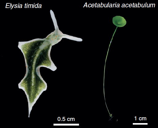
The saccoglossan sea slug, Elysia timida and its primary food source (and plstid contributor) Acetabularia acetabulum.  Image from de Vries et al., 2014

The sacoglossan sea slug, Elysia timida (left) and its primary food source and plastid contributor, Acetabularia acetabulum (right). Image from de Vries et al., 2014

Research by de Vries et al. (2014) examined chloroplast genome content from the algae Acetabularia acetabulum and Vaucheria litorea, the preferred food sources of the sea slugs, Elysia timida and Elysia chlorotica respectively.  This study revealed that the chloroplasts encoded the genes tufA and ftsH, which are not present in the plastids of many land plant chloroplasts.  Furthermore, after a month of starvation, transcripts of the plastid genes tufA, ftsH and psbA were all found in E. timida (de Vries et al., 2014) supporting the previous findings of tufA and ftsH transcripts from V. litorea plastids in E. chlorotica after two months of starvation. The presence of these genes are noteworthy since ftsH is essential for the maintenance of the photosystem II (a major part of the photosynthetic pathway), and tufA plays a role in enabling the protein from fstH to be made. It is also noteworthy that, while the sea slugs eat other algae including Bryopsis sp., the Bryopsis chloroplast is not retained by the sea slugs, and does not encode the ftsH gene.

This could mean that the presence of ftsH, tufA and other genes involved in maintaining the photosynthetic function are the key to long-term retention of algal chloroplasts in sea slugs. It is possible that the sea slugs are not actually providing any protein to the kleptoplast and are not actively maintaining the plastid. With this in mind it seems only logical that we conclude that these sacoglossan sea slugs have evolved into photosynthetic pirates, plundering algae for their plastids and digesting everything else in their wake.

Photosynthetic Sea Slug Pirate. Image from: http://javaape42.deviantart.com/art/Pirate-Slug-colored-380400956


References:

Bhattacharya D, Pelletreau KN, Price DC, Sarver KE, Rumpho ME. (2013) Genome analysis of Elysia chlorotica egg DNA provides no evidence for horizontal gene transfer into the germ line of this kleptoplastic mollusc. Mol Biol Evol 30:1843-1852.

De Vries J, Habicht J, Woehle C, Huang C, Christa G, Wägele H, Nickelsen J, Martin W Gould S. 2014. Is ftsH the key to plstid longevity in sacoglossan sea slugs? Genome Biol Evol 5:2540-2548

Rumpho ME, Pelletreau KN, Moustafa A, Bhattacharya D. 2011. The making of a photosynthetic animal. J Exp Biol 214:303-311.

Wägele H, Deusch O, Händeler K, Martin R, Schmitt V, Christa G, Pinzger B, Gould SB, Dagan T, Klussmann-Kolb, A, Martin W. 2011. Transcriptomic evidence that longevity of acquired plastids in the photosynthetic slugs Elysia timida and Plakobranchus ocellatus does not entail lateral transfer of algal nuclear genes. Mol Biol Evol 28:699–706.

Posted in Uncategorized | Leave a comment

The Bermuda Seaweed Project: Education and Outreach

 As a graduate student studying Bermuda’s seaweed biodiversity, I had the good fortune of spending a year in the islands, collecting thousands of macroalgal specimens for the Bermuda Seaweed Project with support from NSF Award No. 1120652. While diving Bermuda’s spectacular reefs on the hunt for new and interesting species of algae was the primary focus of our work, my colleagues and I were eager to share our enthusiasm for these beautiful and vital organisms with the Bermudian community— especially its students.  For those who aren’t in the know, algae are commonly misconceived as being ‘green’ (some are, but not most!) or ‘pond scum’ (most algae live in the sea, not fresh water) or ‘stinky’ (most only smell bad when they are dead).  Another misconception is that seaweeds are mainly a nuisance— mucking up beaches and overgrowing the more charismatic organisms on a reef— like corals.  But to the contrary, macroalgae create essential habitats for juvenile marine animals, in addition to being important primary producers.  They also provide terrestrial beasts (humans) with a multi-billion dollar industry tied to health and medicine, food production, and cosmetics.  Seaweeds exhibit a staggering diversity of sizes, shapes, colors, textures and habitats, and the exquisiteness of some is an inspiration to biologists and artists alike.  For all these reasons, we find it quite satisfying to share our knowledge of and affection for these underappreciated organisms by creating opportunities for education and outreach.  My work in Bermuda inspired several formal education/outreach activities, which I will describe below, as well as countless informal occasions where I’d like to think my interest in algae gave my peers a fresh perspective on this diverse and important group of marine organisms.

In July and August of 2012, I was a guest educator for the Bermuda Institute of Ocean Science (BIOS) Ocean Academy Marine Science Internship.  This program serves students ages 16 to 18, and focuses on getting kids involved in scientific diving. Education Coordinator Kaitlin Baird and I developed a workshop for student interns that included a reef macroalgae scavenger hunt (via scuba).

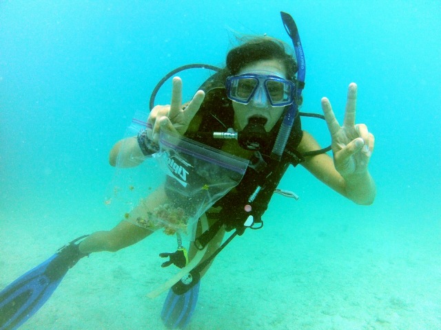
BIOS Ocean Academy student intern collecting algae via scuba!

BIOS Ocean Academy student intern collecting algae via scuba!

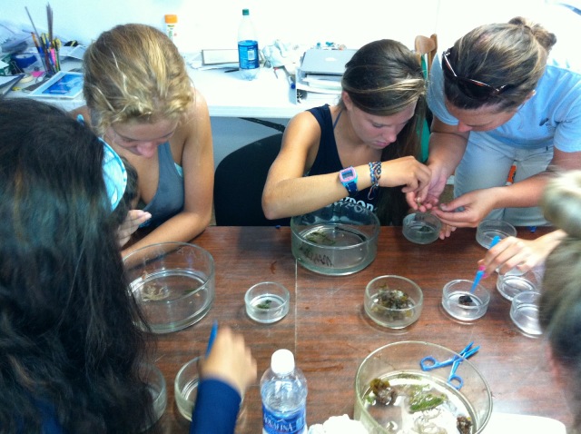
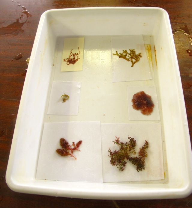
Later I provided guidance to students as they identified specimens, and taught them how to float and press algae to create herbarium records for their collections.

Sorting and identifying algae back in the lab

Sorting and identifying algae back in the lab

As an extension of these lessons, Kaitlin designed some interactive online media to distribute to Bermuda’s teachers that explains how to make an herbarium.  This is a wonderful way to encourage the intersection of science and art!  You can check that out here:

http://prezi.com/buybvbyjpj5u/how-to-make-a-herbarium/

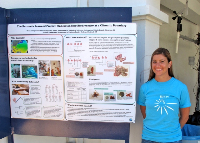
Each year, BIOS hosts an island-wide community event, Marine Science Day, where BIOS research is showcased for the public. Islanders of all ages can view featured presentations, participate in marine-science related activities, and talk to scientists about their work.  I participated in the September 2012 event by sharing an overview of my work in a poster presentation, accompanied by a ‘touch tank’ of some of Bermuda’s most common red, green and brown algae.

BIOS Marine Science Day 2012

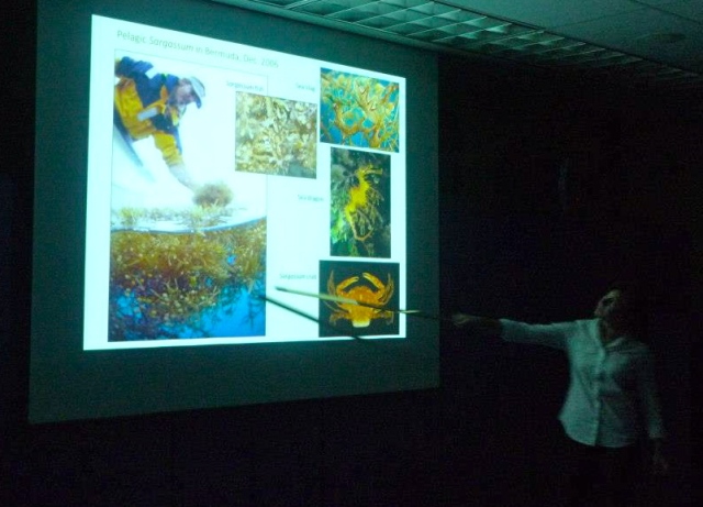
In December 2012, toward the culmination of my field year in Bermuda, I presented my work to a public audience via the Bermuda Aquarium Museum and Zoo’s Lecture Series.  I spoke about the biology and evolution of macroalgae, as well as its importance in the ecosystem, and explained the significance of my research project in the islands with some major findings to date.

BAMZ Lecture Series

BAMZ Lecture Series

Early this year, BIOS conducted their annual Explorer Program— this year’s was ‘Expedition Sargasso,’ and focused on the biology, chemistry and physics of the Sargasso Sea. About a thousand of Bermuda’s primary school students (ages 8-12) visited the event.  Students learned about what algae is, how it is found, identified and pressed. A main highlight was the brown algae of the genus Sargassum.  Students spent time trying to separate out some of the most common species of Sargassum in Bermuda using a dissecting scope. They went on to learn about the wealth of life that calls this seaweed home, along with the movement of the beds within the North Atlantic Gyre, and more specifically the Sargasso Sea.

The event took place under an artful representation of a Sargassum bed.

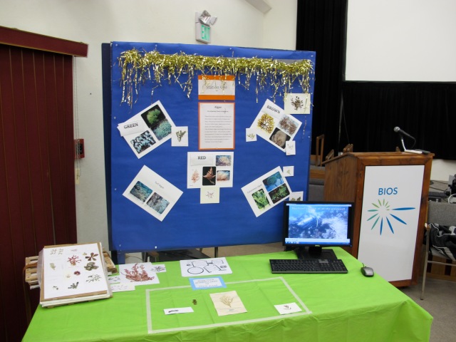
Educators transformed the ceiling of the exhibit space into a floating Sargassum bed, and showcased an algae station where students and teachers could learn about the three major groups of seaweeds, look at herbarium specimens of a variety of Bermuda’s algal species, and watch a video of our team diving for algae on the reef.

Seaweed table with looping video of our team collecting algae while diving!

Seaweed table with looping video of our team collecting algae while diving!

You can watch the hunt here!

Posted in Uncategorized | Tagged , , | Leave a comment

Corals Recruit Gobies as Bodyguards

The Disney movie, Finding Nemo, popularized the understanding that coral reefs provide essential habitats for a large amount of marine biodiversity including hundreds of species of and fish, turtles, invertebrates, and many others.  Unfortunately, over the past thirty years, half of the Great Barrier Reef has been lost and nearly 80% of the live coral in the Caribbean has died.  With such a drastic decrease in the area that serves as feeding, spawning, and nursing grounds for such a wide array of marine organisms, one can only expect increasing competition for territory and a decrease in marine biodiversity.  In a new study, published in the November 9th edition of Science, researchers found that some of the reef inhabitants are helping to fight back against further coral reef loss.

Currently, major stressors including ocean acidification and heating, pollution, disease, and the overfishing of herbivores, enable some seaweeds to increasingly outcompeting the corals for territory.  Corals in the genus Acropora are essential for reef biodiversity as they provide the topography that other species rely on for colonization.  This study found that two reef dwelling gobies, Gobiodon histrio and Paragobiodon echinocephalatus, would help to protect the coral Acrospora nasuta from advances by the toxin containing seaweed Chlorodesmis fatigata.

The authors found that colonies of C. fastigata that wereplaced on corals inhabited by either or both species of gobies were quickly eaten or removed by the fish.  Abundance of the seaweed was decreased by 30% over the course of three days and the damage to A. nasuta was reduced by ~80% in comparison to that in corals lacking the gobies.  Additionally, the seaweed was found in the gut of the goby G. histrio, while it was not found in the gut of P. echinocephalatus.  It is also noted that G. histrio produces a toxic skin secretion, while P. echinocephalatus does not, though further investigation was unable to link the metabolites from the seaweed to the toxins in the goby.  These gobies are providing the coral with a fighting chance as these toxic algae continue to expand their range.

A goby on its way to remove C. fastigata from the coral Acrospora. Photo: Danielle Dixson

Recognition of the mutualistic relationship between the coral and the gobies was not enough, and the authors also set out to see what triggered the goby’s response to the encroaching seaweeds.  Using a cleverly designed experiment the authors found that chemicals released during the interaction between C. fastigata and A. nasuta along with cues from stressed coral after C. fastigata was removed caused the gobies to move toward the site of contact.  However, the seaweed on its own would not alert the gobies.  Further study using faux seaweeds controls and faux seaweeds infused with extracts from C. fastigata provided evidence that when compounds from C. fastigata come in contact with the coral, A. nasuta releases chemical cues, alerting the goby, which then arrive to remove the seaweed from the area.  When experiments were replicated using different coral species in contact with C. fastigata, no response from the gobies was detected.Though similar to other mutualistic relationships that have been well studied, the authors note that this appears to be the first time studies have shown one species chemically triggering a response in another species to help remove a competitor.

Additional research has shown that C. fastigata is particularly damaging to a range of Acrospora species and its continued presence will slow the recovery of corals that have been damaged (Bender et al., 2012).  A greater understanding of chemical cues may lead to other mechanisms that can be employed, slowing the loss of the earths remaining coral reefs.  Until that time comes, hopefully these little gobies can continue to combat C. fastigata, so corals like Acrospora can continue serving as the foundation that allows reefs to flourish and be large centers of biodiversity for generations to come.

References:

Bender, D., Diaz-Pulido, G., and S. Dove. 2012. Effects of macroalgae in corals recovering from disturbance. Journal of Experimental Marion Biology and Ecology. 429: 15-19.

Dixon, D.L., and M.E. Hay. 2012 Corals chemically cue mutualistic fishes to remove competing seaweeds. Science. 338: 804-807.

Posted in Uncategorized | Leave a comment

Aunt Ruby’s Got Good Genes

Few edible plants inspire the nostalgia or controversy that a tomato does; a quick Google search uncovers hundreds of articles and blog posts whose authors covet the tomatoes of yore, and admonish modern day supermarket–bound tomato handling practices like forced ethylene gas ripening, or the abominable, flavor-depleting refrigeration. Few garden staples can lend themselves more to the myriad of possibilities, raw or cooked, sliced, sauced or roasted, salsa-ed or gazpachoed or bloody mary-ed, that a tomato can. In perhaps its finest incarnation, still warm from the summer sun, with only a drizzle of olive oil and a sprinkle of salt, it is, quite literally, the stuff of poetry (see Pablo Neruda’s Ode to the Tomato). For me, it was this same longing, disdain, and appeal that made one genome publication among a plethora of genome publications stand out. The Tomato Genome.

Jason Waskey, Small Heirloom Tomato and Salt, oil on linen, 2009.

Indeed, as reported in Nature this May, an extensive, global team of scientists has successfully sequenced the genomes of the domesticated tomato, Solanum lycopersicum (specifically, the Heinz 1706 cultivar), as well as its closest wild relative, Solanum pimpenellifolium. The massive project has been ongoing since 2003. The information generated from these efforts is of particular significance given the evolutionary history of the Solanum lineage—which is among the world’s most diverse plant genera— and its ability to shed light on the mysteries of eudicot evolution. Also, because tomatoes are such an important food crop, scientists, breeders, farmers, and consumers may benefit from a greater understanding of the molecular and genetic architecture of tomato strains if that knowledge is used to produce superior fruits.

The authors, who are collectively referred to as the “Tomato Genome Consortium,” made two important discoveries when comparing their sequenced tomato genome to the grape genome. First, their data supported the hypothesis a whole-genome triplication occurred in the common eudicot ancestor and second, an additional polyploidy event occurred in the tomato lineage following divergence from grapes, and is shared with the potato genome (Sato et al., 2012). These triplications likely contributed to the major eudicot lineages, and may help to explain, at least partially, what continues to be a hotly debated topic since Darwin reflected on it centuries ago— the rapid diversification of angiosperms on the planet (Stockey et al. 2009).

Figure 2a. from Soto et al. below.

Speciation and polyploidization in eudicot lineages. Confirmed whole-genome duplications and triplications are shown with annotated circles, including ‘T’ (this paper) and previously discovered events α, β, γ. Dashed circles represent one or more suspected polyploidies reported in previous publications that need further support from genome assemblies. Grey branches indicate unpublished genomes. Black and red error bars bracket indicate the likely timings of divergence of major asterid lineages and of ‘T’, respectively.

The result of polyploidy events are the generation of new gene families that, in the case of tomatoes, have had important functions in the plant’s fruit. For example, scientists determined (using orthology and synteny analyses) that gene families mediating functions such as fruit ripening and lycopene (antioxidant) biosynthesis were generated during such genome duplications. Interestingly, these evolutionary outcomes have impacted the loss of fruit functions as well, with important implications for their viability as a food source— gene subfamilies connected to the production of toxic compounds in ancestral lineages have either been completely lost in tomatoes, are not expressed (Sato et al., 2012). Texture is another key characteristic that determines how appetizing a tomato is. Genes sequenced during this project show that cell wall structure and composition largely affect fruit texture, with over fifty differentially-expressed genes encoding proteins involved in altering the structure of the cell wall.

Then how might this extensive data set be applied to the tomato breeding industry? By identifying traits that are associated with fruit development, might we manipulate genetics (through breeding) to select for desirable traits such as taste, texture, color, pest-resistance, speed of ripening, shelf life, pathogen susceptibility, or even levels of antioxidant and disease-fighting phytochemicals?

In a chapter of Genetic Diversity of Plants devoted to the tomato, authors Bauchet and Causse (2012) note that tomato bottlenecks that were artificially generated through domestication, leading to a drastic reduction of genetic diversity, followed by diversification in domestic types by selective breeding. Prized in modern CSA culture, tomato ‘heirlooms’ were pollinated from the seeds farmers saved from the previous year’s harvest; new genotypes arose either from spontaneous mutations, natural hybridization or purposeful recombination of tomato plants with varied traits.

Colorful ‘heirloom’ tomatoes in a variety of shapes and sizes.

A great challenge for breeders has been the ability to ‘unlink’ favorable and unfavorable traits (You say tomato, 2012) i.e. optimal taste and pest attractiveness. Now, scientists can help them conquer this by knowing not only what trait a particular gene codes for, but also how its placement within the genome may influence the expression of neighboring genes. The tomato genome project has generated millions of SNPs (Sato et al., 2012) that can be used  for biodiversity-based breeding, a traditional alternative to the production of genetically-modified “frankenfoods,” which consumers are generally wary of.

Sequence comparison of the Heinz 1706 genes to those of the wild relative, S. pimpenellifolium, revealed that a number of gains and losses of stop codons may have significantly altered gene function in the domesticated tomato. However, Sato et al. found several chromosomal segments in cultivated tomatoes, especially the small “cherry” variety, that are more closely related to the wild species than to Heinz 1706, and attribute this to gene pool mixing through breeding. With both genomes sequenced, greater opportunity arises for new varieties to be cultivated based on wild-type genes, with the potential of reintroducing alleles that were lost during domestication (Bauchet & Causse, 2012). For the likes of farmers, gardeners and foodies everywhere, I hope the resulting traits will render tomatoes as tasty and as healthful as ever.

References:

Bauchet, G. & Causse, M. (2012). Genetic diversity in tomato (Solanum lycopersicum) and its wild relatives Genetic Diversity in Plants DOI: 10.5772/33073

Sato, S. & The Tomato Genome Consortium (2012). The tomato genome sequence provides insights into fleshy fruit evolution Nature DOI: 10.1038/nature11119

Stockey, R.A., Graham, S.W. & Crane. P.R. (2009) Introduction to the Darwin special issue: the abominable mystery. Am. J. Bot. 96: 3-4.

Anon. (2012). You say tomato Nature DOI: 10.1038/485547a

Posted in Uncategorized | Leave a comment

Microdissection with frikin laser beams!

ResearchBlogging.org
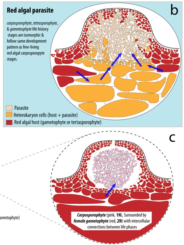
One of the problems we have to deal with when approaching some of the parasite work we do is figuring out how to access small patches of tissue. With the red algal parasites, this is particularly true, because of the way they grow. Many of the cells that contain parasite DNA also harbor host DNA, because they are heterokaryon cells (Top of Figure 1).

Figure 1. Comparison of parasite life history and the carposporophyte stage of a typical red algal life history. Parasite cells devoid of host DNA occur only in a portion of the “erumpent pustule” that extends from the host. Image from Blouin & Lane 2012 (DOI:10.1002/bies201100139)

If one wants to work with the genomes of these organisms, it is essential to be able to identify and extract tissue that contains only parasite DNA, which proves to be a challenge. We have played with several methods without success… until now. Postdoc Nic Blouin has been work with the Rhode Island Children’s Hospital, using their laser capture microdissection (LCM) system to isolate red algal cells of interest.

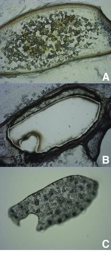
With our parasites living on the West Coast, we had to devise a proof of concept with a local species. We picked the carposporophytes (bottom of Figure 1) of Chodrus crispus as a test case, based on their size similarity to many red algal parasites. Nic isolated the carposporophytes, dried and sectioned them in prep for LCM (Figure 2A).

Figure 2. Carposporopyte tissues was dried and sectioned using a freezing microtome. A) Thin sections were mounted for LCM. B) After cutting, the C) cystocarp material was excised from the carposporophyte.

Nic was able to use the UV laser to cut out the cystocarps (Fig. 2B) and then, using transfer film, was able to remove the excised cells (Fig 2C). An explanatory video of the capture process can be found here. Removal of the cystocarp cells is roughly equivalent to the size and nature of the parasite material we will be isolating in the near future. From here we plan to use multiple isolates (to avoid reaction biases) and whole genome amplification to produce enough DNA to create a library for Illumina sequencing. We should have a feel for how much DNA we can produce in a week or two.

References:
Blouin NA, & Lane CE (2012). Red algal parasites: models for a life history evolution that leaves photosynthesis behind again and again. BioEssays : news and reviews in molecular, cellular and developmental biology, 34 (3), 226-35 PMID: 22247039

Posted in Uncategorized | Tagged , , , | Leave a comment

To Beard or Not to Beard. That is the Question.

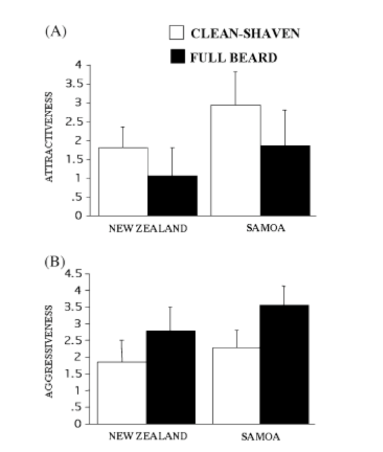
You are a man standing in front of the mirror. Do you go for the razor or do you skip the morning shave? Well, it should depend on how you want women to perceive you. According to Dixson and Vasey (2012), if you keep that beard you will be observed as older, of higher social status, and more aggressive. On the other hand, if you take the time to shave that beard, you are going to convey attractiveness to women.

Facial hair is a sexual dimorphism that appears in male children around the time of puberty. Beard hair continues to grow until maturity and it is notably thicker and coarser than hair anywhere else on the head. While testosterone plays a role in beard growth, ethnicity is more a more important factor in determining the pattern, distribution, and density of the beard. Women do not have beards and this indicates it is not retained by natural selection (thermoregulation/UV protection). A more likely reason for facial hair in men is sexual selection.

Intrasexual selection leads to the appearance of traits that signal competitive ability. Under this mode of selection, a beard would signal confidence and competitive ability. Based on studies of lions, a beard does not offer any protective advantage from blows or attacks.  It has been suggested that a beard is a hazard during fights or could harbor parasites (Zahavi & Zahavi 1997). This could also provide women with an indirect signal of fitness (strength/immunity).

Beards have been shown to convey traits favorable to females including: masculinity, maturity, confidence and age. This intersexual selection for beards is not strongly supported in humans. Among the primates, many species have sexual adornments around the face for display purposes. It is hypothesized that intersexual selection, or female choosiness, is likely the driving force behind beard evolution. Most previous facial hair studies have focused on Western cultures and lack the strength of cross-cultural comparison.  The participants in this study are from different socioeconomic backgrounds and different exposure to mass media.

Figure 1: Participants were recruited with full beards, defined as not having shaved or trimmed the face for at least 6 weeks. The men are from New Zealand and Samoa. These men were photographed with a full beard and then again when cleanshaven posing smiling (A), angry (B), and neutral (C) facial expressions (from Dixson and Vasey 2012).

The photographs were presented individually, in a random order and rated using a 6-point Likert Scale, where 0 = unattractive, 1 = only slightly attractive, 2 = moderately attractive, 3 = attractive, 4 = very attractive, and 5 = extremely attractive.  The same scale was used using the terms aggressive and important. Both women and men were asked to give their perceived age of the men photographed. For the New Zealand sample, participants were of European descent and rated only the images of men who were of European descent. Samoan participants rated only the photographs of Samoan men.

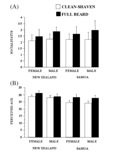
Figure 2: (A) Mean social status scores. Men and women from both cultures rated men with beards as more important. (B) Mean perceived age. Actual mean age is 23 years for both cultures.

These study results suggest that the beard plays a stronger role in intrasexual signaling than in female mate preferences. In this study, they tested the extent to which facial expressions and beards act in concert in the perceptions aggression. Dixson and Vasey found beardedness did enhance the perception of male dominance, elevated social status, and the communication of aggressive intent. Previous studies have found beards decrease a man’s perceived social status due to its association with vagrancy (Morris 2002); however, participants in this study ascribe a higher social status to bearded males. Whether or not the beard reliably advertises men’s actual strength or is associated with greater reproductive success (like other masculine traits) remains to be determined.

Figure 3: (A) Mean attractiveness scores made by women. Both cultures rated clean-shaven as more attractive, but Samoan women had higher ratings for clean-shaven (B) Mean aggressiveness scores made by women. Both cultures rated bearded as more aggressive.

In addition to beards, androgens drive other sexually dimorphic traits such as a large jaw, narrow eyes, a pronounced brow ridge, and a longer face. Men displaying these masculine traits are ranked as less ‘‘warm,’’ honest and cooperative, and less trustworthy (Dixson & Vasey 2012). Darwin postulated that beards evolved in human ancestors via female choice as a highly attractive masculine adornment. Facial hair has varied in popularity and style, and while this study removed the element of Western influence it still found bearded men less attractive to women. These results in concert with previous studies may prove Darwin wrong or simply that female choice is highly variable.

Now that the facts are known, the choice to shave the beard or let it grow is up to the individual. Beards may convey the perception of wealth, status, or age, but when it comes down to the basics, the whole goal of existence for any species is reproduction and women just don’t dig the beard. And gentlemen, that beard is going to hurt your chances.

Works Cited

Dixson BJ, Dixson AF, Morgan B, Anderson MJ. 2012. Beards augment perceptions of men’s age, social status, and aggressiveness, but not attractiveness. Behavioral Ecology. Online Adv Access.

Morris D. 2002. People watching. London: Vintage.

Zahavi A, Zahavi A. 1997. The handicap principle. Oxford: Oxford University Press.

Posted in Uncategorized | Leave a comment

Oh What a Big If

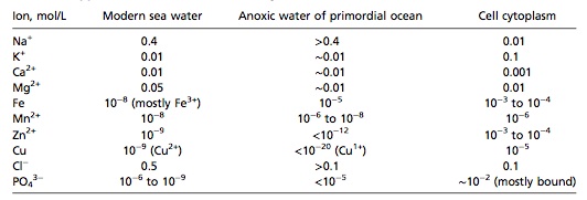
A team of scientists led by Armen Y. Mulkidjanian at the University of Osnabrück in Germany have invoked Charles Darwin’s “warm little pond” scenario to challenge one of the most prevalent views held in evolutionary biology— that life originated in the sea, not on land.  Using geochemical and phylogenomic data for support, their primary argument stems from the idea that the chemical environment of the ocean, even at hydrothermal vents, is too dissimilar to that of the internal cellular environment to have sustained life in the early stages of cell evolution. The authors contend that the chemistry of protocells would have been similar to that of the environment in which they were created, because these cells had not evolved sufficiently to contain sophisticated membrane barriers and pumps to compensate for disequilibrium between their internal and external environments.  In searching for environments on Earth that might better represent the chemical composition of cells, they discovered that the chemistry of condensed geothermal vapor forming pools at inland geothermal fields were a fit, especially with respect to the high K+ to Na+ ratio found in cells (and opposite to that present in seawater).

"Warm little ponds" at Yellowstone National Park.

Mulkidjanian et al. also present an alternative hypothesis— that the differences between the chemistry of the cell and of the marine environment are not related to the conditions in place which early cells were evolving, and that the cell’s requirements for certain ions evolved later, after transmembrane pumps allowed cells to exist in disequilibrium.  The authors reject this hypothesis however, on the inference that the most ancient proteins common to life require specific ions (e.g. potassium, zinc, phosphate, etc.) in concentrations that are predicted at the described geothermal pools.  Particularly interesting from a phylogenomic perspective, data analysis showed that the origin of GTPases (involved in protein synthesis) preceded the last universal common ancestor (LUCA).  These have highly conserved K+ binding sites, suggesting that K+ was required early in the evolution of life.  Additionally, the authors note that phosphates (which form the backbones of nucleic acids) are “four orders of magnitude greater than in seawater” in the cytoplasm of the cell.

Approximate total concentrations of key ions in modern sea water, the primordial ocean, and cell cytoplasm.

Several assumptions must be made for the arguments of Mulkidjanian et al. to hold up. The evidence presented is only acceptable if one presumes cells have not undergone dramatic changes to their chemical composition in the last 4 billion years of life (a notion that isn’t too hard to swallow given that the cells of all extant organisms use the same basic chemistry to function), if one believes the chemistry of modern cells should reflect the chemistry of the primordial environment where cells first evolved (what Mulkidjanian et al. cite as “the chemistry conservation principle”) and if the chemistry of the ancient sea (especially at hydrothermal vents) had not significantly changed since the early evolution of life, hence reflecting the low K+/Na+ ratios found in modern marine environments.  The authors point to a reference supporting the similarity of Archaean seawater to modern oceans, but do not elaborate on this conjecture.

With such a complicated and long debated topic as the origin of life, it goes without saying that a study such as the present one will generate quite a bit of controversy.  My initial evaluation upon reading the paper included being impressed and intrigued by the authors’ novel ideas and thorough reasoning, but I was also eager to read the refutations that were sure to have followed its publication.  Indeed, articles in Nature, Scientific American, and presumably in other reputable science and news publications offered commentary from some prominent scientists who study the origins of life.  Challenges included the unlikelihood that biological molecules could have remained in such unstable and transient environments as volcanic pools for a long enough period to evolve (Switek, 2012), that direct evidence of the described environments would be nearly impossible to ascertain from the fossil record, and that the acidity of these geothermal pools would have been too great to allow life (though this point is addressed by Mulkidjanian et al., who maintain that pH would have been closer to neutral in the absence of oxygen on the primordial Earth).  Though, some, like molecular biologist Jack Szostak of Harvard Medical School, agreed that protocells undeniably had leaky membranes, rendering the early ocean a hostile environment for cells due to its high levels of sodium (Biello, 2012).  However, Szostak does not agree that the K+/Na+ ratio of internal cells necessarily reflects the early environment.  It may simply be that cells functioned better under such ion concentrations, and thus selection for high K+ followed early evolution (Switek, 2012).  Perhaps future research might aim to elucidate the mechanisms by which leaky protocells may have maintained these favorable ionic conditions internally, despite the incompatible chemistry of the external environment.

References:

Biello, D.  February 15, 2012.  Did life’s first cells evolve in geothermal pools?  Scientific American.  Retrieved from http://www.scientificamerican.com/article.cfm?id=did-life-first-evolve-in-geothermal-pools

Mulkidjanian, A. Y., Bychkov, A. Y., Dibrova, D. V., Galperin, M. Y. & Koonin, E. V. (2012) Origin of first cells at terrestrial, anoxic geothermal fields.  Proc. Natl Acad. Sci. USA advance online publication http://dx.doi.org/10.1073/pnas.1117774109.

Switek, Brian.  February 13, 2012.  Debate bubbles over origin of life.  Nature News.  Retrieved from http://www.nature.com/news/debate-bubbles-over-the-origin-of-life-1.10024

Posted in Uncategorized | Leave a comment

The Table Ate My Dinner

My thoughts after reading Gerber et al (2012) situations like that may not be science fiction for ever.  Inspired by natural living surfaces, such as bark or our own skin, the authors set out to create a functional living material that was capable of self cleaning.  Using PVC, agar incapasulated fungi (Penicillium roqueforti), and a 0.4 micron polycarbonate surface membrane (Figure 1) they were able to create a living surface capaable of “eating” a food spill.  The key to the succes of this material is it’s simplicity.  With a total thickness of 300 microns and the a shelf life of at least 10 days this conseptual design shows extreame promise to the devolepment of living surfaces.  The key to this technology is the surface membrane which is porous enough to allow for macromolecule exchange while have pores small enough to prevent the fungal hyphae from escaping their contained layer.  This serves to prevent both the fungi and the outside from contamination.

Figure 1. "Design of a living surface. The simplest form of a living surface is composed of three layers. The base layer (light blue) may be an inert support foil (e.g. a PVC film), a linker, or a surface that shall be coated directly with a living surface. The second layer is the living layer hosting microorganisms. The top layer (black, wireframe) is responsible for the confinement of the fungi, for nutrient, gas, and product transportation, and mechanical, chemical, and biological stability..."

A few of the success that I found most interesting was the ability for this material to respond to a variety of realistic cleaning situations, its localized response to spills, and it’s ability to clean multiple spills.  Gerber et al used ethanol hand sanitizer and hand soap on the surface of the material to mimic typical surface cleaning procedures.  In both cases sterilizing effects were limited to the outer surface of the membrane, as the biological response of the fungi to a food spill was unaffected post cleaning.  The material also displayed localized responses to food.  By creating isolated food spill on the same surface the authors demonstrated that the fungi would only proliferate at the site of the spill while other areas of the surface remained dormant.  Finally, they were able to generate multiple cleaning cycles from the same mat.  Food was introduced at two time points over a 30 day period, both instances of the spill were met with the same fungal response and resulting self cleaning.  Fascinating.

The success of this conceptual model allows us to speculate about other applications of this novel technology.  Changing the biological portion of the membrane can allow for a variety of other uses.  By incorporating antibiotic, or toxin, producing microorganisms this technology can be used to produce site and dose specific responses to bacterial populations. Truly self-sterilizing surfaces. This application alone represents a multi-million dollar area of research.  Since the response of the biological organism could simply be a visual que, surfaces could be designed to identify contamination sites at very early stages.  Applications of living materials extend to packaging, air quality, numerous consumer products, and laboratory settings.

As with all novel technologies the grand possibilities are used to overshadow the obvious problems.  In the case of the functional surface presented here I see multiple issues that would prevent this from attaining a production level item.  First, the shelf life while seemingly manageable at 10 days is masked by the fact that the foil must remain wet for that entire time period.  Failure to keep the foil moist resulted in a null response to a food spill.  The cost of storage may outweigh the actual benefits of the use.  Second, while this foil does “eat” or “self-clean” it does so at a snails pace.  It takes 8 days for the foil to absorb 2 mL of a glucose solution.  Not a timeline my wife would approve of for cleaning up a spill.  Lastly,  the membrane which allows for macromolecule transfer and keeps the fungi “trapped” is destined to become ruptured in any consumer usage.  Before a production product could even be discussed the surface membrane would have to be replaced with something that could handle the realistic wear and tear of a consumer product.

While a fascinating and promising endeavor into functional biological surfaces many more years of R & D will be required before we have a viable consumer product.  I guess stories of dinner eating tables will remain science fiction for now.

Gerber, L., Koehler, F., Grass, R., & Stark, W. (2011). From the Cover: Incorporating microorganisms into polymer layers provides bioinspired functional living materials Proceedings of the National Academy of Sciences, 109 (1), 90-94 DOI: 10.1073/pnas.1115381109

Posted in Uncategorized | Tagged , | Leave a comment

When a tree isn’t actually a tree.

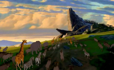
Elton John may have first introduced the idea in his hit song, Circle of Life, but a similar phrase has found its way into science. Undoubtedly Elton John considered only charismatic megafauna in his song, but if applied to the three Kingdoms of life, a circular evolutionary model arises. It is my suspicion that the authors Rivera and Lake tossed around the term “Circle of Life” during the preparation of their manuscript, but wisely chose against The Lion King reference. The Ring of Life idea was first brought to light in 2004, but still appears in textbooks and is widely cited, thus it deserves some revisiting.

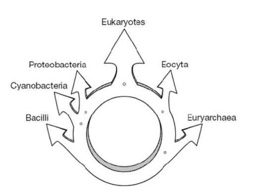
Figure 1: Since the time of Darwin, the Tree of Life has been used to represent the evolutionary relationships among species. However, a more accurate representation may be a Circle of Life (Elton John, The Lion King), a Web of Life (Bapteste & Walsh 2005), or a Ring of Life (Rivera & Lake 2004).

Lake and Rivera pioneered a novel new algorithm based on gene content, called conditoned reconstruction, for genome based tree construction. This approach relies on the analogy gene:genome as nucleotide:gene. During evolution the orthologous gene can become either present (P) or absent (A). Thus a genome alignment can occur and Markov models can be applied to reconstruct the evolutionary history. Because genome comparison across all domains of life is impossible,  Lake and Rivera introduce a ‘conditioning genome’ to serve as a reference to compare genomes. This type of analysis can detect the fusion of two genomes opposed to the several large lateral gene transfers.

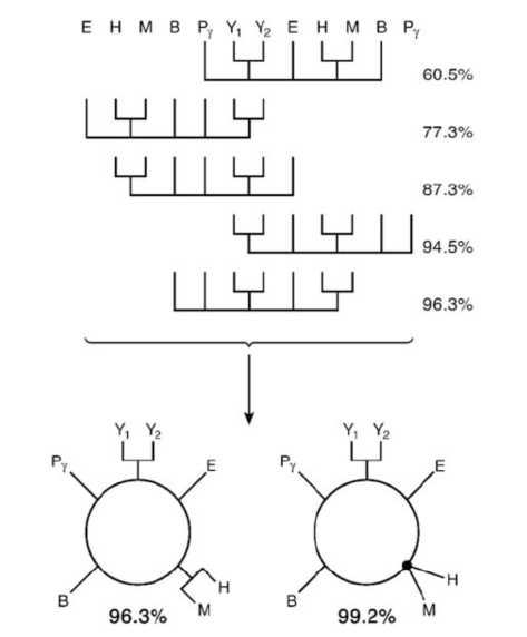
Rivera and Lake applied this model to eight genomes (3 Bacteria, 3 Archea, and 2 Eukaryotes). The resulting 5 trees are shown in Figure 2. The cumulative probabilites of these trees is shown at the right. The unrooted trees are shifted so a repeating pattern is revealed which indicates that the trees are actually variations of an underlying cyclic pattern. The rings shown at the bottom of Figure 2 are the compilation of the 5 trees, and when cut at any of the 5 arcs and unraveled, the resulting tree is one of the original 5 trees.

Figure 2. The 5 most probable trees in order of decreasing bootstrap support. From top to bottom: 60.5%, 16.8%, 10%, 7.2%, and 1.8% support. Fully resolved ring is at left and partially resolved ring is on the right. Results are based on 2,408 orthologousgene sets. From Rivera and Lake (2004).

These results strongly support the conclusion that 2 prokaryotic genomes fused, resulting in the first eukaryote. Rivera and Lake conclude the eukaryotic nuclear genome formed from the fusion of a proteobacterium and an archael eocyte (see Fig 3). Because a fusion organism connects two nodes of a ring, the removal of the fusion organisms will open the ring and convert it into a tree. The ring opened only when the 2 eukaryotes (yeast) were removed which indicates that the yeast lineage is the product of prokaryotes. Rivera and Lake also add to the evidence that in the Eukaryotic genome, informational genes derived from Archea and operational genes from Bacteria during the two genome fusion event.

Figure 3. Ring of Life schematic. The Tree of Life is indeed more ring-like than trunk-like according to the analysis of Rivera and Lake. The eukaryote resulted from the fusion of an archaen (eocyta and euryarchae) and a bacteria (proteobacteria, cyanobacteria, and bacilli). From Rivera and Lake (2004).

If coroborated, this Ring of Life provides a signifiicant departure from a long accepted evolutionary theory. In a subsequent paper, Bapteste and Walsh  (2005) claim that Rivera and Lake misrepresent lateral (horizontal) gene transfer and provide criticm of the methods. Since 2005 the Ring of Life has fallen out of favor, but many scientists believe it may be true and it just needs adequate support. As Elton John sings, “In the Circle of Life/ There’s far to much to take in here/ More to find that can ever be found”. The Tree of Life has been ever evolving and much debated, but leave it up to scientists, the answer will be found.

Works Cited:

Lake, J.A. and Rivera, M.C. (2004) Deriving the genomic tree of life in the presence of horizontal gene transfer: conditioned reconstruction. Mol. Biol. Evol. 21, 681–690

Rivera, M.C. and Lake, J.A. (2004) The ring of life provides evidence for a genome fusion origin of eukaryotes. Nature 431, 152–155

Bapteste, E. and Walsh, D.A. (2005) Does the ‘Ring of Life’ ring true? Trends Microbiol, 13, 256-261

Posted in Uncategorized | Leave a comment

Biological NAND Gate: The future may be closer than we think

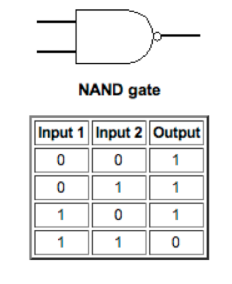
Digital circuits are built out of seven simple logic gates:  AND, OR, XOR, NOT, NAND, NOR, and XNOR.  These logic gates work by receiving a simple input and producing an output based on the input.  The AND gate will return a ‘True’ Boolean value if and only if both of its inputs are true, while the NOT gate will return a Boolean value of ‘True’ if the input is ‘False’, just like the words would suggest. Biologists at the Imperial College London have constructed an AND Gate and a NOT gate and were able to link them to produce a combitorial NAND Gate.  The NAND gate will return ‘True’ if both of the inputs are not ‘True’ (Figure 1).

Truth table of a NAND Gate. (http://whatis.techtarget.com/definition/0,,si_ gci213512,00.html)

The biological NAND gate (Figure 2) was constructed in E. coli.  The inputs are outputs are all natural biological molecules.  There are two inputs that go into the AND gate.  If both of the inputs are ‘True’, it will pass a ‘True’ value onto the NOT gate, which will then return a ‘False’.  If either of the inputs are ‘False’, the result of the NAND gate will be ‘True’.

Diagram of the synthetic NAND gate (Wang et al 2011).

Biological logic gates are not completely novel, but these new gates are able to be used in a number of biological systems and are modular, meaning that they can be joined together.  These improvements could lead to faster development of more complex biological circuits.  Once the seven simple logic gates are able to be constructed we will be able to make biological computing as complex as we would like.  Martin Buck, one of the authors on the paper, suggested that the gates could form the building blocks of microscopic biological devices.  His ideas include: sensors that can swim inside of arteries, search for harmful plaque, and deliver medications; sensors that could detect and destroy cancer cells inside the body; and sensors that could be deployed into the environment to detect and neutralize dangerous toxins (Quick, 2011).  They also propose that advanced circuits could be built to work as a projected image edge detector or a cellular event counter (Wang et al, 2011).  The possibilities are literally endless, and the future of medicine may be closer than we think.

Quick, D. (2011, October 21). Logic gates created from dna and bacteria could form basis of biological computers. Gizmag, Retrieved from http://www.gizmag.com/biological-logic-gates/20237/

Wang, B. et al. Engineering modular and orthogonal genetic logic gates for robust digital-like synthetic biology. Nat. Commun. 2:508 doi: 10.1038/ncomms1516 (2011).

Posted in Uncategorized | Leave a comment